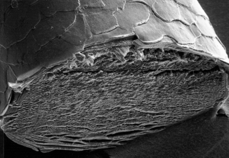
Examining hair under a microscope is a fascinating science investigation. Students can use low-power stereo microscopes to study various types of strands under observation to learn to differentiate them based on color and shape differences. hair strands are flat scales made of the fibrous protein known as keratin. Their protection comes from an outer layer called the cuticle, which, under microscope examination, looks similar to an unfurled sock.
Compound Microscope
A compound microscope is a light microscope that enables users to observe tiny objects. Utilizing multiple lenses to magnify them, its magnification powers can reach 1000x. It can be used for studying plants, bacteria, and blood smear samples, among many other things. Objective lenses can be found in the nosepiece of a microscope and adjusted to produce various magnification levels, typically 4X to 100X. They usually feature threaded mounts, so additional objectives may be added anytime. Once your sample is in position, carefully set it on a slide and secure it with stage clips. Adjusting the coarse focus knob until your specimen is centered within its aperture or stage opening allows you to use the fine focus knob to fine-tune its position until its focus at maximum magnification is necessary for viewing it.
Stereo Microscope
Stereo or dissecting microscopes are excellent tools for studying opaque samples, using light that reflects off them to illuminate them. They can be enhanced further with additional lenses to alter operating distance and magnification levels; other lenses may even help correct image distortion. Compound microscopes use separate objective lenses and eyepieces for each eye to create three-dimensional images of specimens, using lower magnification but providing excellent resolution at high detail levels. Stereo microscopes can be used for many purposes, from sorting to dissecting. Some popular features of stereo microscopes are their apochromatic optics, darkfield imaging capabilities, high magnification range of 10x to 50x magnification range, and replaceable lenses. Stereo microscopes can also be mounted onto boom stands, articulating/flex arms, plain frames, track stands, table mounts, and illumination systems for optimal viewing experience.
Scale Casting
Scale casting is an invaluable technique used in hair examination laboratories, enabling examiners to see the pattern of scales on specimens when they have dried or become tacky. A thin medium layer must be spread onto a glass microscope slide before pressing hair fiber against it. Once dry, this cast can be pulled off, leaving behind an impression or model of cuticular scales visible through its pores. Oxidizing metals exposed to water vapor lead to thicker oxide scales than those seen on clean surfaces due to the rapid diffusion of oxygen atoms through its surface and carbon accumulation in its composition. Furthermore, oxygen from steel substrate can permeate through cracks in the oxide scale, affecting its design further.
Photomicrographs
Photomicrographs are taken through a microscope with an attached camera and then saved or printed as still images for further processing. With digital cameras becoming increasingly accessible and affordable, photomicrography has grown increasingly popular. Metal and stone objects may be ground, chemically etched, and mounted onto slides for photographic examination under a metallurgical microscope. On the other hand, biological samples can be killed, stained, and mounted for photographic observation under transmitted light under an ordinary light microscope or different types of microscopy such as fluorescence, infrared, electron, or X-ray microscopy. Photomicrographing is an intricate form of photography requiring extensive knowledge of microscope and camera techniques. Furthermore, illumination under a microscope tends to be lower than traditional daylight or flash photography, and using filters helps mitigate reciprocity failure.